1 Introduction In 1988, a Brazilian scholar M. worked in the Research Group of the Department of Physics of the University of Paris, France. N. Baibich discovered the giant magnetoresistance (GMR) effect when studying the electronic transport properties of Fe/Cr magnetic superlattice thin films. That is, the resistivity of the material changes significantly with the magnetization state of the material.
This discovery has attracted the attention of scientists in many countries. The basic research and applied research of the giant magnetoresistance effect and its materials have quickly become a hot topic. Since then, the research on the giant magnetoresistance effect has developed rapidly and has been based on Research and applied research have gone hand in hand and have become an international paradigm for rapidly transforming basic research into commercial applications. Currently, GMR materials have been commercially used in fields such as magnetic sensors, computer read heads, and magnetic random access memories.
The sensor made of GMR material is called a giant magneto-resistive sensor. It has the advantages of high sensitivity, wide detection range and resistance to harsh environments. It can utilize semiconductor exposure and etching processes to make the device integrated and miniaturized, and its cost performance is far Superior to other kinds of magnetic field sensors This paper reviews a new type of sensor combining GMR sensor and biotechnology--GMR biosensor. This sensor is applied to the field of biological detection and is a sensor for detecting magnetically labeled biological samples. The immune magnetic microsphere (IMB), high magnetic sensitivity GMR sensor and associated readout circuit are composed of three parts.
2 Immunomagnetic Microspheres In 1979, John Ugelstad et al. successfully prepared a polystyrene microsphere with appropriate uniformity and particle size. After being magnetized and linked with an antibody, it became an immune system with excellent separation effect. Magnetic labeling - dynabeads Since then, immunomagnetic labeling has been widely used and has led to a revolution in biological separation technology. The characteristics of immunomagnetic labels are fast separation, high efficiency, good reproducibility, simple operation, and no expensive Equipment, biological properties and functions that do not affect isolated cells or other biological materials.
Immunomagnetic microspheres, or immunomagnetic labels, are magnetic microspheres with monoclonal antibodies on the surface. It is a new immunological technique that has been popular in recent years at home and abroad. It is based on immunology and penetrates into pathology and physiology. In various fields such as pharmacology, microbiology, biochemistry, and molecular genetics, its applications are widely used, especially in immunological detection, cell separation, protein purification, etc. There are now many companies in the world that specialize in the production of magnetic labeling products. For example, Dynal, Nanomag, Micromer and other domestic companies that produce this product include Ningbo Xinzhi Biotechnology Co., Ltd., Hangzhou Lianke Biotechnology Co., Ltd., Shenzhen Nano Microbial Technology Co., Ltd., etc. Figure 1 shows some immunomagnetic labels. The magnetization curve, of which Dynal's M-280 is the most commonly used immunomagnetic marker for GMR biosensor detection, diameter 2.8μm, with superparamagnetism. 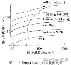
The basic principle of immunomagnetic labeling technology is as follows: Immunomagnetic labels can bind both active proteins (antibodies) and magnets. After a certain treatment, antibodies can be bound to magnetic labels to make them the carriers of antibodies. After binding of the antibody on the label to the specific antigen, an antigen-antibody-magnetic labeling immune complex is formed. The functional group of the immunomagnetic label is mainly bound to the protein, but the immunomagnetic label can also be made by the avidin-biotin system. And non-protein binding, such as various DNA, RNA molecules. In order to make the immune magnetic marker play a greater role.
3 High-sensitivity GMR sensors At present, there are mainly four types of magnetically ordered materials with GMR effect, which are derived from experiments and theoretical studies: multilayer film structures, spin valve structures, magnetic alloy particle structures, and particle-film composite structures. The structure has its own characteristics, and GMR biosensors mostly use multilayer membrane structures or spin valve structures.
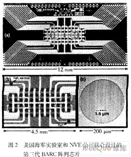
In 1998, the US Naval Laboratory took the initiative to use the GMR effect and immunomagnetic labeling to realize the idea of ​​a GMR fetal sensor. They tested the feasibility of the principle by measuring DNA, antigen-antibody, donor and acceptor experiments. The Magnetic Tag Array Counter (BARC) was further proposed, and a DNA array chip was developed. Figure 2 is a third-generation BARC array chip jointly designed by the US Navy Navy Lab and NVE Corporation. The planar layout is shown in Figure 2(a). Fig. 2(b) is a partial enlargement of Fig. 2(a). It uses a semiconductor process to integrate a 64-channel GMR sensor on a silicon substrate. Each sensor consists of a total length of 8 mm and a width of 1.6 μm. The ground is distributed in a circular area with a diameter of 200 μm (Fig. 2(c)). The magnetoresistance value is 42 kΩ, the saturation magnetization and GMR effect (ΔR/R) are 30 mT and 15%, respectively, for each sensor. A multi-layer film structure in which the detection sensor uses a magnetic layer/a non-magnetic layer/a magnetic layer can be separately performed, and the two magnetic layers separated by the non-magnetic layer are anti-parallel coupled.
In addition to the U.S. Naval Laboratory and NVE Corporation, Stanford University in the United States, the University of Bielefeld in Germany, and the University of Lisbon in Portugal also conducted research on GMR biosensors. In China, the Institute of Electrical Engineering of the Chinese Academy of Sciences, the Institute of Electrical Engineering, Chinese Academy of Sciences, Although Tsinghua University and the University of Electronic Science and Technology have made some progress, they lack the organic integration of biotechnology and their development is relatively backward.
GMR sensor detection process shown in Figure 3, first, in the sensor surface for the specific detection of biological probe (Figure 3 (a)), and then make the test solution flow sensor surface, the specific target molecules in the test solution will be The probe captures (Figure 3(b)) and then adds the immunomagnetic microspheres. The immunomagnetic microspheres interact with the target molecule to complete the label (Figure 3(c)). In this case, an applied gradient magnetic field perpendicular to the sensor surface is required. Excess immunomagnetic microspheres that do not participate in label separation can reduce the background noise during detection, thereby improving the detection accuracy. Then, the magnetic labels are magnetized by an applied alternating magnetic field, and the magnetic alternating magnetic labels generate additional alternating energy. The magnetic field induces a change in the sensor's magnetoresistance. By reading the change in the magnetoresistance, it can determine whether there is a target molecule in the test solution to be detected, and determine the concentration of the target molecule in the test solution according to the magnitude of the change in the magnetoresistance.

4 signal detection circuit magnetoresistance changes need to be converted into electrical signals, there are two ways to achieve, one is the Wheatstone bridge structure, as shown in Figure 4 (a), the other is the use of IV conversion method, as shown in Figure 4 (b) shows.
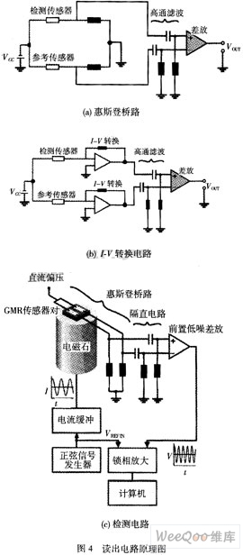
Both output signals remove the background noise represented by the reference signal in the detection signal and then amplify it. However, the noise due to the physical reasons of the material and the device cannot be completely eliminated. When the detection signal is very weak, The signal-to-noise ratio is too low, the above circuit can not realize the readout of the signal. At this time, the phase-locked amplification technology must be used to read out the signal. The detection process is shown in Fig. 4(c). The phase-locked amplification technique is used for the weak signal. One of the effective methods of detection is to use a cross-correlation technique to amplify and detect signals in the signal under test that are synchronized with the reference signal.
The lock-in amplifier consists of three parts: the signal channel, the reference channel and the correlator (also called the phase detector). The function of the signal channel is to amplify the weak signal to a level high enough to drive the correlator, and it also has the suppression and filtering part. The function of interference and noise; Correlator is a unit circuit that completes the cross-correlation function calculation of the measured signal and the reference signal, and is composed of a multiplier and an integrator circuit; the reference channel provides a periodic signal with the same frequency as the measured signal.
At present, the signal detection of GMR biosensors is based on common general-purpose lock-in amplifiers on the market, and its full-scale sensitivity can reach the order of nV. However, most of them are modular test instruments, which are too large and expensive to be used. For the marketization of products, it is very necessary to design a GMR sensor chip and semiconductor technology with good compatibility, and it can be packaged together with the lock-in amplifier IC chip using MCM technology, which will greatly improve the GMR biosensor The practicality and popularity.
5 Conclusion In summary, the giant magnetoresistance biosensor integrated technology, semiconductor technology, magnetic thin film technology and weak signal detection technology in one, through the detection of immunomagnetic markers, can accurately determine the composition of the test solution and contained The concentration of components, etc., is a successful extension of the GMR sensor in the field of biological detection because of its many advantages such as high sensitivity, high resolution, low cost, miniaturization of equipment, and automation of measurement processes, etc., in life sciences, medicine, and defense, etc. The application potential in the field is huge, and with the advancement of semiconductor technology, its degree of integration and sensitivity will be further improved. However, at present, research on GMR biosensors is still at the basic stage of research at home and abroad. There is still a certain distance from practicality.
DH650 Dry Roll Press Granulator
Dry Roll Press Granulator Co., Ltd. , http://www.nspelletmill.com